LINKEDIN.COM
RxSight, Inc. posted on LinkedIn
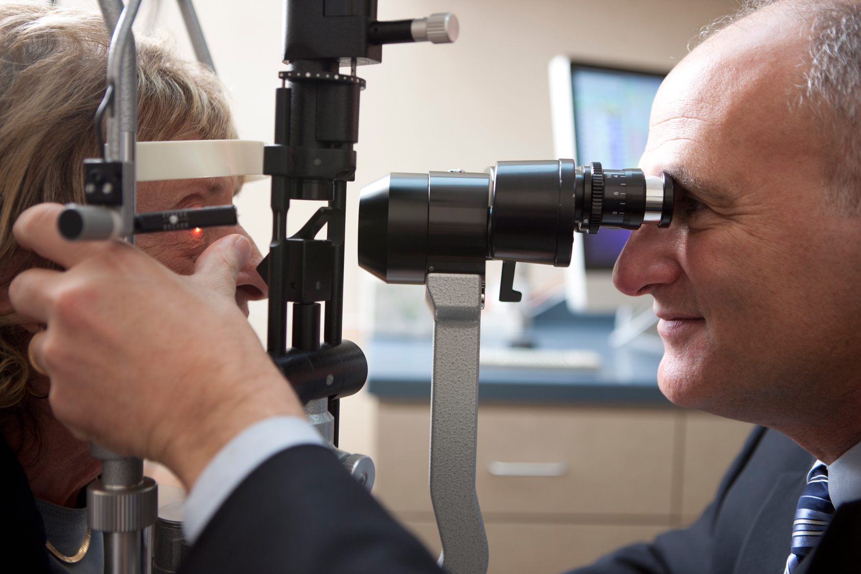
Photos from our office.
Our practice will start seeing patients again on Monday, May 11th in full compliance with all local, state, and federal guidelines. This includes masks, social distancing, and the disinfection of all common surfaces. Elective surgeries are also being rescheduled. Care will be taken to avoid office congestion and minimize exposure to COVID-19.
We require: ➡️ You do not have fever, chills, shaking, muscle pain, headache, sore throat, or a loss of taste or smell ➡️ You have not had exposure to anyone with a flu-like illness within the past two weeks ➡️ You wear a mask or equivalent facial covering over both your nose and mouth ➡️ You agree to maintain social distancing ➡️ You avoid touching your eyes, nose, mouth, and face ➡️ You enter the office alone if possible
Expect to hear from our staff shortly. You may contact us at your convenience from links at jameslewismd.com
Chorioretinal folds are a known finding following penetrating glaucoma surgery, as in these two cases who underwent Ahmed valve tube shunt placement. Prevalence is estimated between 10-50% of incisional glaucoma surgeries.
Pic 1.) several linear chorioretinal folds throughout the posterior pole. Intraocular pressure was 4mmHg at the time of this photo. The fundus and visual acuity returned to baseline within a week as IOP leveled at 10mmHg.
Pic 2.) Small choroidal folds can be seen distributed temporal to the macula. This image also demonstrates a large hemorrhage consistent with ocular decompression retinopathy.
Pic 3.) shows complete resolution of the choroidal folds and hemorrhage after 4 weeks in patient 2.
#ophthalmology #ophthalmologist #ophthalmictech #ophthalmologyresident #ophthalmicphotography #ophthalmicsurgery #cornea #corneasurgery #eyesurgeon #eyesurgery #eye #oculardisease #optometry #optometrystudent #optom #sunyoptometry #osuopt #salusuniversity #glaucoma #glaucomasurgery #retina #chorioretinalfolds @ Lewis LASIK
Inflammation of the anterior chamber can create fibrin plaques that are readily seen within the pupil. The second and third images demonstrate an almost completely occluded pupil with synechia formation. The fourth image demonstrates an ultrasound biomicroscopy image of a patient in angle closure following complete pupil occlusion from fibrin (blue arrow). Aggressive corticosteroid therapy can ‘melt’ the fibrin and cycloplegics can mechanically disrupt it. Nd:YAG laser can also instantly disrupt total occlusion.
#ophthalmology #ophthalmologist #ophthalmictech #ophthalmologyresident #ophthalmicphotography #ophthalmicsurgery #cornea #corneasurgery #eyesurgeon #eyesurgery #eye #oculardisease #optometry #ocularinflammation #optometrystudent #optom #sunyoptometry #osuopt #salusuniversity @LewisLASIK
Descemet stripping endothelial keratoplasty (DSEK) is a corneal transplant procedure that replaces only the innermost cells of the cornea. It is readily combined with cataract surgery to improve refractive outcomes. This is a one day post operative visit of a DSEK showing faint edema and remaining air bubble. The air bubble will typically dissolve over the first 48-72 hours.
#ophthalmology #ophthalmologist #ophthalmictech #ophthalmologyresident #ophthalmicphotography #ophthalmicsurgery #cornea #corneasurgery #eyesurgeon #eyesurgery #corneatransplant #dsek #fuchsdystrophy #endothelium #eye #oculardisease #optometry #optometrystudent #optom #sunyoptometry #osuopt #salusuniversity
Lewis LASIK updated their address.
